Blood Vessels Labeled / Blood Vessel Structure And Function Boundless Anatomy And Physiology - The blood vessels are the components of the circulatory system that transport blood throughout the human body.
Blood Vessels Labeled / Blood Vessel Structure And Function Boundless Anatomy And Physiology - The blood vessels are the components of the circulatory system that transport blood throughout the human body.. Blood vessels are flexible tubes that carry blood, associated oxygen, nutrients, water, and hormones throughout the body. Blood vessels (labeled) coloring page. 10 photos of the the human blood vessels labeled. They all have a small smooth inner layer of called the endothelium. • identification of blood vessels as arteries, capillaries or veins from the structure of their walls.
The iliac, femoral, popliteal and tibial (calf). They all have a small smooth inner layer of called the endothelium. Pictures and 3d models played a great role in helping me learn. If a blood vessel breaks, tears, or is cut, blood leaks out, causing bleeding. Label the blood vessels and structures using the hints provided.
Histology Of Blood Vessels from www2.victoriacollege.edu Blood vessel segmentation in retinal fundus images. 10 photos of the the human blood vessels labeled. This page provides histology support information for blood vessel structure. They also take waste and carbon dioxide away from the tissues. Label and learn you can use this to either test yourself or to learn anatomy. Does not cover the pathology content. Blood, the heart and the vessels that carry blood around the body together make up the cardiovascular system. Blood vessels are vital for the body and play a key role in diabetes helping to transport glucose and insulin.
What do you mean by the term.
Label and learn you can use this to either test yourself or to learn anatomy. As a medical student, i found anatomy pretty challenging. This is an online quiz called blood vessel labeling. Blood vessels 2 labeled palmar arch digital artery right femoral a right femoral v great saphenous vein left popliteal a right anterior tibial a. Abstract—the segmentation of retinal blood vessels in the retina is a critical step in diagnosis of diabetic retinopathy. An extraordinary degree of branching of blood vessels exists within the human. Carry blood away from the heart (always oxygenated apart from the pulmonary artery which goes from the heart to the lungs). The difference in the structural characteristics of arteries, capillaries and veins is attributable to their. Partial barrier resistance is labeled resistant and total resistance is labeled proof. Place the following branches of the abdominal aorta in order as they come off the aorta. Label the blood vessels and structures using the hints provided. Hma practical 3 for monday july 23 and wednesday july 25. Does not cover the pathology content.
Does not cover the pathology content. Blood vessels (labeled) coloring page. Three types of blood vessels that make up the entire system. Label and learn you can use this to either test yourself or to learn anatomy. Does not form part of the actual practical class based upon the virtual slides.
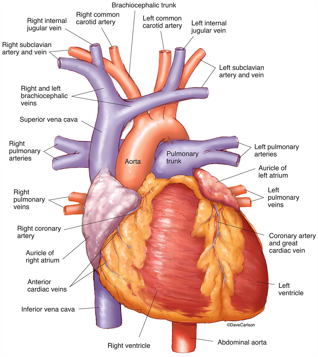
Diagram Of Heart Blood Vessels Auto Electrical Wiring Diagram from www.carlsonstockart.com This page provides histology support information for blood vessel structure. Blood vessels 2 labeled palmar arch digital artery right femoral a right femoral v great saphenous vein left popliteal a right anterior tibial a. Blood vessels can be damaged by the effects of high blood glucose levels and this can in. Blood vessel segmentation in retinal fundus images. B.mention three adaptation of plant to reduce excessive transplration. These vessels transport blood cells, nutrients, and oxygen to the tissues of the body. Arteries, arterioles, capillaries, venules, and veins. What do you mean by the term.
These vessels transport blood cells, nutrients, and oxygen to the tissues of the body.
Greisler, in principles of tissue engineering (fourth edition), 2014. The iliac, femoral, popliteal and tibial (calf). Pictures and 3d models played a great role in helping me learn. The blood vessels are the components of the circulatory system that transport blood throughout the human body. Does not form part of the actual practical class based upon the virtual slides. 10 photos of the the human blood vessels labeled. Partial barrier resistance is labeled resistant and total resistance is labeled proof. They are vital for carrying nutrients, oxygen and waste around the body. Blood vessels 2 labeled palmar arch digital artery right femoral a right femoral v great saphenous vein left popliteal a right anterior tibial a. Blood may flow out of the body, as external bleeding, or it may flow into the spaces around organs or directly into organs. • identification of blood vessels as arteries, capillaries or veins from the structure of their walls. As a medical student, i found anatomy pretty challenging. Blood vessels are vital for the body and play a key role in diabetes helping to transport glucose and insulin.
Label the veins of the upper limb. If a blood vessel breaks, tears, or is cut, blood leaks out, causing bleeding. 2,731 blood vessels labeling machine products are offered for sale by suppliers on alibaba.com, of which labeling machines accounts for 5. All blood vessels have some features in common. 10 photos of the the human blood vessels labeled.
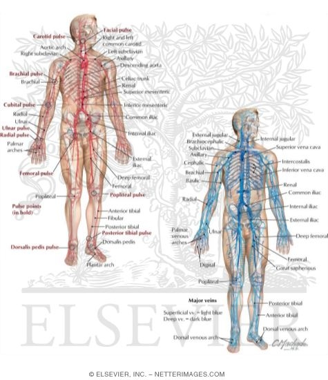
Major Arteries Pulse Points And Veins from www.netterimages.com As a medical student, i found anatomy pretty challenging. Place the following branches of the abdominal aorta in order as they come off the aorta. Arteries carry blood away from the heart. Blood may flow out of the body, as external bleeding, or it may flow into the spaces around organs or directly into organs. Blood vessels are vital for the body and play a key role in diabetes helping to transport glucose and insulin. They also take waste and carbon dioxide away from the tissues. Does not form part of the actual practical class based upon the virtual slides. Pictures and 3d models played a great role in helping me learn.
• identification of blood vessels as arteries, capillaries or veins from the structure of their walls.
Abstract—the segmentation of retinal blood vessels in the retina is a critical step in diagnosis of diabetic retinopathy. 4.which blood vessel will have the high amount of glucose and amino acld after a meal? Blood vessels can be damaged by the effects of high blood glucose levels and this can in. Blood vessel segmentation in retinal fundus images. Label the veins of the upper limb. Arteries carry blood away from the heart. As a medical student, i found anatomy pretty challenging. Deep veins, located in the center of the leg near the leg bones, are enclosed by muscle. Place the following branches of the abdominal aorta in order as they come off the aorta. Greisler, in principles of tissue engineering (fourth edition), 2014. Veins (in blue) are the blood vessels that return blood to the heart. Vessels labeled diagram, blood vessels labeling exercises, cat blood vessels labeled, human anatomy blood vessels, human blood. Pictures and 3d models played a great role in helping me learn.